
registry : 8(391)220-15-54
reception: 8(391)228-07-47
660022, Krasnoyarsk, st. Partizan Zheleznyak, 3d;
Electron transmission microscopy
A complex research method, when very thin tissue fragments are examined in a special way by the method of electron beam transmission.
The electron microscope makes it possible to distinguish very small objects with a size of only a few nanometers (the size of objects is less than the size of virus particles!).
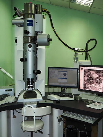
The Bureau uses one of the most modern German-made electron microscopes Libra 120 (Zeiss). At present, there is extensive experience in the use of the electron microscope for diagnosing kidney diseases.
Head of the laboratory of electron microscopy, pathologist of the highest qualification category Gappoev Stanislav Vitalievich.
The range of pathologies in which electron microscopy is used is extremely diverse:
Study of kidney biopsy specimens in the diagnosis of glomerulopathies of various etiologies;
Examination of kidney tissue when planning kidney transplant operations, both in relation to the recipient and the donor;
Skeletal muscle biopsies for suspected mitochondrial pathology, storage diseases, systemic diseases;
Peripheral nerve biopsies in patients with suspected neuropathy or demyelinating disease and more.
The Bureau is the only medical institution in the Krasnoyarsk Territory, the Republic of Tuva and the Republic of Khakassia, which performs this complex research method.
Bureau employees constantly improve their knowledge in this area by participating in seminars, conferences and on-the-job training in leading Russian institutions.
TO THE PATIENT
Who orders this study?
electron microscopy may be assigned:
- directly by the pathologist who conducted the study of your material;
- by your doctor
What is needed to conduct research?
This information is available on our website under "What do you need to analyze"
If you have questions about this type of study, please contact us at 8-923-274-3784. Be sure to leave your contact details so that we can respond to you.

